31/08/2009
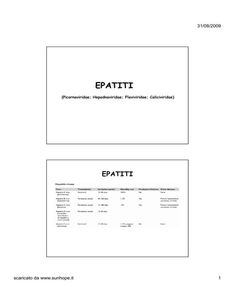
EPATITI
(Picornaviridae; Hepadnaviridae; Flaviviridae; Caliciviridae)
EPATITI
scaricato da www.sunhope.it
1
31/08/2009
…ma vari altri virus possono causare
epatite
•
•
•
•
•
•
•
•
•
CMV
EBV
HSV
VZV
Adenovirus
Virus della Rosolia
Virus della Parotite
Virus delle febbri emorragiche
Enterovirus (cox e echo)
scaricato da www.sunhope.it
2
31/08/2009
EPATITI
EPATITI
scaricato da www.sunhope.it
3
31/08/2009
Virus dell’epatite A (HAV)
Hepatitis A virus (HAV) particles observed by immune electron microscopy.
Virus-like particles, 27 to 32 nm in diameter, are arranged in a crystalline
array, with antibody coating their surfaces. These HAV particles appear to
have cubic symmetry and have capsomeres measuring 8 to 12 nm
Phylogenetic relationship of hepatitis A virus (HAV) and simian HAV
(SHAV) to representative species of the mammalian picornavirus genera
scaricato da www.sunhope.it
4
31/08/2009
¾Non-Enveloped
¾Simmetria icosaedrica
¾+RNA
¾Baltimore Class.: IV
Virus appartenente alla famiglia Picornaviridae
scaricato da www.sunhope.it
5
31/08/2009
Ciclo replicativo
1.
2.
Il virus si lega al recettore cellulare
Sito e meccanismo di scapsulamento
ignoti
3. La proteina VPg (pallino arancione)
viene rimossa e l’RNA si associa ai
ribosomi
4. Inizia la traduzione e viene prodotta
una poliproteina
5. Taglio proteolitico della poliproteina
6. Le proteine che partecipano alla
sintesi dell’RNA virale vengono
trasportate a delle vescicole
7. La sintesi dell’RNA avviene sulla
superficie di queste vescicole dove è
anche stato trasportato l’RNA+
8
8.
Avviene la sintesi del filamento
negativo
9. Il filamento – serve come stampo per
la sintesi di quello +
10. Le proteine strutturali vengono
utilizzate nella fase di assemblaggio
11. insieme all’RNA genomico
12. Per uscire infine dalla cellula mediante
lisi cellulare
10
11
12
Acute viral hepatitis. Lobular disarray
characterized by anisocytosis, anisonucleosis,
ballooning, and Kupffer cell hypertrophy
Acute viral hepatitis. Ballooned hepatocytes
show lysis of cell membranes and nuclei and an
empty cytoplasm
Acute viral hepatitis. Two sinusoidal acidophilic
(apoptotic) bodies can be seen in the center of
the field
scaricato da www.sunhope.it
6
31/08/2009
Acute viral hepatitis. Portal area infiltrated
with numerous inflammatory cells
Acute viral hepatitis with submassive necrosis.
Zones of necrosis are linked together (bridging
necrosis)
Acute viral hepatitis with massive necrosis. All
liver cells have “dropped out.” Note
proliferating periportal cholangioles and
infiltration of stroma and portal areas with
inflammatory cells
Immunologic and clinically relevant biologic events associated with
hepatitis A virus (HAV) infection in humans
scaricato da www.sunhope.it
7
31/08/2009
Rates of reported hepatitis A cases (per 100,000 population) in the United
States, by race/ethnicity, 1997
scaricato da www.sunhope.it
8
31/08/2009
Distribution of viral hepatitis cases in the United States reported to
the Centers for Disease Control and Prevention in 1998
scaricato da www.sunhope.it
9
31/08/2009
Virus dell’epatite B (HBV)
scaricato da www.sunhope.it
10
31/08/2009
Virus con envelope
Vi
l
Simmetria icosaedrica
Dna circolare parzialmente bicatenario
(filamento lungo DNA - ; filamento corto DNA +)
Baltimore Class.: I
Il filamento corto si completa solo nell’imminenza della replica del DNA virale
scaricato da www.sunhope.it
11
31/08/2009
DNA polimerasi
presente nel virione
DNA parentale
circolare e
parzialmente
p
bicatenario
RNA polimerasi
cellulare
DNA circolare
bicatenario
superspiralizzato
( + ) RNA
pre-genomico
Replicazione HBV
RNA messaggero
Varie proteine
funzionali
Transcriptasi inversa
DNA polimerasi
DNA della progenie virale
Progenie virale
scaricato da www.sunhope.it
12
31/08/2009
Ciclo replicativo
Pathology of chronic hepatitis B
A: Necrosis (Arrowheads indicate
lymphocytic infiltrates disrupting the
limiting plate of the portal area and
invading the parenchyma)
B Lymphoid
B:
L
h id aggregates.
t
(Arrowhead
A
h d
shows a dense lymphoid aggregate in
the portal area surrounding the bile
duct)
C: Lobular inflammation. Scattered
apoptotic hepatocytes (arrowheads)
with surrounding lymphocytes
D: Cirrhosis. Thick bands of fibrotic
tissues
(arrowhead)
encircling
regenerative nodules
E: Ground-glass hepatocytes
F: Fibrosing cholestatic hepatitis.
Diffuse ballooning degeneration of
hepatocytes
scaricato da www.sunhope.it
13
31/08/2009
scaricato da www.sunhope.it
14
31/08/2009
Serologic and clinical patterns observed during acute HBV infection.
scaricato da www.sunhope.it
15
31/08/2009
scaricato da www.sunhope.it
16
31/08/2009
scaricato da www.sunhope.it
17
31/08/2009
scaricato da www.sunhope.it
18
31/08/2009
Natural history and growth of HCC in HBsAg-positive carriers.
scaricato da www.sunhope.it
19
31/08/2009
Virus dell’epatite C (HCV)
Acido nucleico
Simmetria del capside
Envelope
Struttura del genoma
Classificazione di Baltimore
Polimerasi virale
Diametro
D
m
del virione (nm)
( m)
Dimensione del genoma (kb)
scaricato da www.sunhope.it
RNA
Icosaedrico
+
ss (+)
IV
40-50
10
20
31/08/2009
Altri virus appartenenti alla famiglia Flaviviridae
Flaviviridae
scaricato da www.sunhope.it
21
31/08/2009
Flaviviridae
VIRUS DELL’EPATITE C
FLAVIVIRUS
HCV
virioni rotondeggianti, 40-50 nm di diametro
RNA ( + ) monocatenario lineare di 10 Kb
con pericapside
replicazione citoplasmatica
Ac anti HCV
Trasmissione parentale
Incubazione
b i
20-40
20 0 gg
Molto probabile la latentizzazione
AG nel fegato
mesi
INF
Fino al 1990 (anno di introduzione dello screening) l’ HCV era la causa del
60-90% di epatiti PT.
scaricato da www.sunhope.it
22
31/08/2009
GENOTIPI di HCV
HCV è un virus molto eterogeneo perché molto mutante
•Sei
Sei genotipi maggiori, sulla base dell
dell’analisi
analisi filogenetica delle
regioni del core, E1 e NS5
•Ogni genotipo è suddiviso in sottotipi minori
•Altro tipo di variabilità, le quasi-specie, cioè varianti
spontanee o indotte dalla terapia e presenti nel singolo paziente
infetto
In Italia i genotipi più frequentemente trasmessi sono il
genotipo 1 e il genotipo 3, essendo il genotipo 1 quello più
rappresentato nella nostra popolazione e il genotipo 3 quello
selezionatosi più recentemente nei giovani.
Estimated incidence of acute hepatitis C virus infections in the United States
between 1982 and 1996
scaricato da www.sunhope.it
23
31/08/2009
Proportion of acute hepatitis C cases related to selected risk factors in
the United States between 1983 and 1996. Before testing, transfusion
accounted for about 20% of acute non-A, non-B hepatitis C cases. At
present, transfusion associated hepatitis C has been virtually eliminated
Representation of clinical responses in patients after infection in acute (A)
and chronic (B) hepatitis C
scaricato da www.sunhope.it
24
31/08/2009
Typical histologic features of chronic hepatitis C
A: Portal area with a lymphoid aggregate
B: Portal area showing necrosis.
necrosis The edge of the portal area is disrupted by lymphocytic infiltration.
infiltration
Within the portal area is a lymphoid aggregate surrounding a duct that shows reactive epithelial
changes. This “duct injury” is termed a Poulsen lesion. It is not associated with chronic cholestasis or
duct loss C: Lobular inflammation. Small foci of inflammation are seen within the hepatic parenchyma
in chronic hepatitis C
D: Steatosis. Accumulation of intracellular lipid within hepatocytes is a common feature in chronic
hepatitis C
E: Periportal fibrosis. The earliest fibrotic change in hepatitis C is an expansion of the portal area by
fibrosis. This gives the portal area a stellate appearance
F: Advanced fibrosis. As the disease progresses, more scarring takes place.
Al mondo oltre 80 milioni di donne HCV positive sono in età fertile.
In Italia --Æ prevalenza dell’ Infezione da HCV in gravidanza
1-2,4 %
Nel 40-50% dei casi non è possibile individuare nessun fattore di rischio,
negli altri casi i fattori di rischio sono quelli tipici delle infezioni da HCV:
• 30-40% stupefacenti per via endovenosa
• < 20% trasfusioni di sangue / emoderivati
partner di persona anti HCV positivo
tipo di lavoro svolto
Il rischio di trasmissione verticale dell’infezione da HCV è circa del 6%
Nessun genotipo di HCV è specificamente correlato alla
trasmissione verticale.
scaricato da www.sunhope.it
25
31/08/2009
The relative proportion of hepatocellular carcinoma cases associated with
chronic hepatitis B virus (HBV) or hepatitis C virus (HCV) infections in
different countries. Some parts of the world still have relatively high numbers
of HBV carriers, resulting in a high proportion of hepatocellular carcinoma HCC
cases due to HBV. In other countries such as Japan, HBV is coming under
control resulting in a higher proportion of HCC cases related to HCV.
scaricato da www.sunhope.it
26
31/08/2009
Location on the HCV polyprotein of recombinant antigens and peptides used in
the Ortho enzyme-linked immunosorbent assay (ELISA) (Ortho-Clinical
Diagnostics, Rochester, NY) and the Chiron recombinant immunoblot assay
(RIBA) antibody test systems (Chiron Corporation, Emeryville, CA).
Recombinant antigens are expressed in yeast as fusion proteins with
superoxide dismutase; this protein tag is represented by the hatched area.
scaricato da www.sunhope.it
27
31/08/2009
Virus dell’epatite D
Electron micrograph of the hepatitis delta virus
Hepatitis D (Delta) Virus
δ antigen
HBsAg
RNA
scaricato da www.sunhope.it
28
31/08/2009
HBV – HDV Coinfection
Typical Serologic Course
Symptoms
ALT Elevated
Titre
anti-HBs
IgM anti-HDV
HDV RNA
HBsAg
Total anti-HDV
Time after Exposure
HBV - HDV Superinfection
Typical Serologic Course
Jaundice
Symptoms
Titre
Total anti-HDV
ALT
HDV RNA
HBsAg
IgM anti-HDV
Time after Exposure
scaricato da www.sunhope.it
29
31/08/2009
Structural proteins of the hepatitis delta virus
Western blot of serum-derived
HDAg with a human monoclonal
antibody to HDAg. Extracts of
HDAg from sera of five humans
infected with HDV (lanes 2
through 6), two chimpanzees
(lanes 8 and 9), and an HDVinfected woodchuck (lane 11)
were analyzed. Lane 1 contains
an extract of an HDV-negative
human serum; lane 7, an HDVnegative
g
chimpanzee
p
serum; and
lane 10, an HDV-negative
woodchuck serum. Lane M
contains
molecular-weight
markers, and the relative
molecular masses in kilodaltons
are indicated on the left margin
scaricato da www.sunhope.it
30
31/08/2009
Replication scheme of hepatitis delta virus.
1: Replication begins with
transcription of genomic
RNA encoding the short
form of HDAg (HDAg-S).
Two types of antigenomic
RNAs are produced: an
mRNA that encodes HDAg,
and a greater-than-unitlength antigenome.
The latter undergoes autocatalytic cleavage via
an internal ribozyme and is ligated by a host
enzyme to yield circular antigenomes, which then
serve as template for genome synthesis by a
similar mechanism
mechanism. HDAg
HDAg-S
S is required for RNA
replication.
2: The adenosine within the stop codon for HDAg-S is deaminated to inosine in
approximately 30% of antigenomic RNAs by a host enzyme. 3: During subsequent
RNA transcription, this change appears in the mRNA, which now encodes the long
form of HDAg (HDAg-L), which inhibits RNA replication. 4: The C-terminus of
HDAg-L is prenylated by a host farnesyltransferase, facilitating interaction with
the envelope protein (HBsAg) and subsequent virion formation
Typical morula cells from a patient with hepatitis caused by HDV
scaricato da www.sunhope.it
31
31/08/2009
The serologic patterns of type D hepatitis: Co-infection and superinfection
Top: Coexistent acute hepatitis B and hepatitis D
Middle: Acute hepatitis D superimposed on a chronic hepatitis B virus
infection
Bottom: Acute hepatitis D progressing to chronic hepatitis, superimposed on a
chronic hepatitis B virus infection
The worldwide distribution of HDV infection, as measured by the prevalence
of anti-HDAg in HBsAg-positive individuals with acute or chronic hepatitis
scaricato da www.sunhope.it
32
31/08/2009
Geographic distribution of HDV genotypes
Circles on the map correspond approximately to geographic origin of isolates.
Filled circles, genotype I
horizontal stripes, genotype II
vertical stripes, genotype III.
VIRUS DELL’EPATITE E
CALICIVIRUS
HEV
virus con forma isometrica, 35-40 nm diametro
RNA lineare ( + ) monocatenario
sprovvisto di peplos
replicazione citoplasmatica
•
•
•
•
trasmessa attraverso il circuito oro – fecale (acqua contaminata)
incubazione più lunga di HAV
le epidemie colpiscono varie migliaia di persone
presente in Asia centrale, USSR, Algeria, Africa centrale, America latina,
Borneo, ……..
• i colpiti sono gli adulti 15-40 anni
• alta mortalità nelle donne in gravidanza
diagnosi: esclusione delle altre forme più dati epidemiologici.
scaricato da www.sunhope.it
33
31/08/2009
L’HEV è attualmente stato spostato dalla
classificazione come Calicivirus ed è tornato ad
essere considerato tra gli agenti virali ad RNA
non classificati.
Acido nucleico
Simmetria del capside
Envelope
Struttura del genoma
Classificazione di Baltimore
Polimerasi virale
Diametro del virione (nm)
Dimensione del genoma (kb)
RNA
Icosaedrica
ss (+)
IV
35-40
8
Fotografia al microscopio
elettronico
l tt
i d
dell virus
i
dell’epatite E
scaricato da www.sunhope.it
34
31/08/2009
In ogni caso la famiglia virale a cui sembra essere più vicino
rimane quella dei Caliciviridae
CALICIVIRIDAE
scaricato da www.sunhope.it
35
31/08/2009
CALICIVIRIDAE
Hep E Genome organisation
Starting from the 5´ end of ORF 1 and extending to its 3´ end, motifs
characteristic of
f the f
following
g viral p
proteins have been identified
f
(55):
¾
A methyl transferase, consistent with the finding that the 5´ end
of the viral genome is capped (42)
¾
A Y domain, a sequence of unknown function found in certain other
viruses, including rubella virus
¾
A papain-like cysteine protease, a type of protease found
predominantly in alphaviruses and rubella virus (35)
¾
A proline-rich “hinge region” that is thought to impart flexibility to
the molecule and that contains a region of hypervariable sequence
(82 94)
(82,94)
¾
An X domain of unknown function that flanks papain-like protease
domains in the polyproteins of other positive-strand RNA viruses
(35)
¾
A domain containing helicase-like motifs that are most closely
related to the helicases of superfamily I (34)
¾
The motifs of an RNA-dependent RNA polymerase, most closely
related to supergroup III of viral RNA polymerases (54)
scaricato da www.sunhope.it
36
31/08/2009
Hepatitis E Virus Infection
Typical Serologic Course
Symptoms
IgG anti
anti-HEV
HEV
ALT
Titer
IgM anti-HEV
Virus in stool
0
1
2
3
4
5
6
7
8
9
1
0
1
1
1
2
1
3
Weeks after Exposure
scaricato da www.sunhope.it
37
31/08/2009
Algorithm for the serologic diagnosis of HAV, HBV, HCV, and HDV.
scaricato da www.sunhope.it
38

