ISOLATION AND CHARACTERIZATION OF
PORCINE CIRCOVIRUS TYPE 2 (PCV2) IN WILD
BOARS IN CENTRAL ITALY
GAVAUDAN S.1, BAROCCI S.1, BRISCOLINI S.1, MANCINI P.1, MORANDI F.3,
PANICCIA’
PANICCIA’ M.1, FERRARI M.2, VILLA R.2, PETRINI S.
S.1
1 Istituto
2 Istituto
Zooprofilattico Sperimentale dell’Umbria e delle Marche, Perugia, Italy;
Zooprofilattico Sperimentale della Lombardia e dell’Emilia-Romagna, Brescia, Italy;
3 Università degli Studi di Bologna, Bologna, Italy.
RESULTS
INTRODUCTION
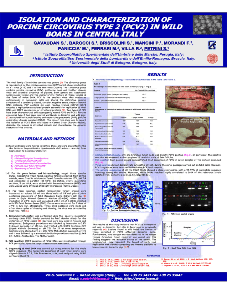
Necropsy and histopathology: The results are summarized in the Table
Table 1 and Table 2.
The viral family Circoviridae contains two genera (1). The Gyrovirus genus
is represented by the chicken anemia virus (CAV) which shows similarities
similarities
to TT virus (TTV) and TTVTTV-like mini virus (TLMV). The Circovirus genus
contains porcine circovirus (PCV), psittacine beak and feather disease
disease
virus and Columbid circovirus of pigeons. Both genera are icosahedral
icosahedral
nonenveloped virions and the characteristic feature of these viruses
viruses is
the form of virion DNA. PCV is the smallest virus that replicates
replicates
autonomously in mammalian cells and shares the distinctive genome
genome
structure of a covalently closed, circular, negative sense, single
single--stranded
DNA molecule. PCV contains six open reading frames (ORFs); ORF1
encodes a replicationreplication-associated protein essential for replication of viral
DNA and ORF2 encodes major structural proteins (2). Two types of PCV
have been characterized and subsequently named PCV1 and PCV2.
PCV2. Porcine
circovirus type 2 has been isolated worldwide in domestic and wild
wild pigs,
(7) associated with proliferating and necrotizing pneumonia (PNP), porcine
respiratory disease complex (PRDC). The aim of this study was to
to report
the isolation of PCV2 from wild boars in Central Italy (Marche Region),
Region),
describe the lesions in affected animals and characterize the genomic
genomic
features of the isolates.
MATERIALS AND METHODS
Sixteen wild boars were hunted in Central Italy, and were presented
presented to the
the Istituto Zooprofilattico Sperimentale dell’
dell’Umbria - Marche from
September to December 2006 for:
1)
2)
3)
4)
5)
6)
Necropsy;
Histopathological investigations;
Virological investigations;
Immunohistochemical examination;
PCR and Real timetime-PCR tests;
Sequencing of viral DNA.
1.1.-2. For the gross lesions and histopathology,
histopathology, target tissue samples
(lungs, mediastinic lymphlymph-nodes, palatine tonsils) collected from all the
animals, were fixed in calciumcalcium-buffered formalin solution 10%, pH 7.4
and embedded in paraffin (56(56-58°
58°C, BioBio-Optica, Italy). Microtome
sections, 5 µm thick, were stained with haematoxylinhaematoxylin-eosin (H&E), and
were viewed using Olympus IX51 light microscope (Tokyo, Japan),
Table 1
Macroscopic lesions detected in wild boars at necropsy (Fig.1
(Fig.1 – Fig.2)
Fig.2)
Organs
Bronchial lymph nodes (enlarged
enlarged and pallor)
16 / 7
Lungs (apical
apical pneumonias with haemorrhages)
16 / 4
Tonsils (Punctiform
Punctiform haemorrhages)
haemorrhages
16 / 1
Table 2
Prevalence of histological lesions in tissue of wild boars with infection by
PCV2.
Tissue
Lesions
Tonsils
5. PCR reaction: ORF2 sequence of PCV2 DNA was investigated through
PCR protocols (3) on the target tissues above mentioned.
6. Sequencing of viral DNA was carried out using primers for the whole
ORF2 sequence. The nucleotide sequences of each virus isolate were
were
aligned (Bioedit 7.0.9, Ibis Biosciences, USA) and analyzed using
using NCBI
software (BLAST).
Lymph nodes
Lungs
Coagulative necrosis
16/6a
16/1
16/0
Hyperplasia of lymph nodes
N.A.
16/2
N.A.
Haemorrhagic Necrosis and
Hemorragiae
16/1
16/1
16/0
Granulomatous inflammation
16/0
16/3
16/0
Giant multinucleate cells
16/1
16/1
16/1
Peribronchial mononuclear
Infiltrates
N.A.
N.A.
16/1
Fibrous bronchiolitis
N.A.
N.A.
16/1
Intracytoplasmatic inclusion
bodies
16/0
16/1
16/0
Fig.2 – Enlarged and pallor of the
Bronchial lymph nodes
a
No. animals tested / No. animals positive
N.A., Not Applicable
Immunohistochemically,
Immunohistochemically, only one bronchial lymph node was slightly PCV2 positive (Fig.3).
(Fig.3). In particular, the positive
reaction was observed in the cytoplasm of dendritic cells of few follicles.
PCR reaction from pooled organs demonstrated DNA sequences of PCV2 in seven samples of the sixteen examined
(Fig. 4).
Virus isolation did not demonstrate cytopathic effect, during the serial passages
passages carried out on NSK cells. However,
PCV2 sequences were detected by RealReal-time PCR in all samples.
samples.
The genome sequencing of all the PCV2 DNAs showed a close relationship, with a 9595-97% of nucleotide sequence
homology among the strains. Moreover, these DNAs resulted highly correlated to DNA of the reference strain
isolated from domestic pigs (Acc. Nr. DQ346683).
1 2 Fig.
3 44 –5 PCR
6 7
9 10 11 12 13 14
pool8 organs
481 bp
3.3.-5. For
For virus isolation,
isolation, pooled homogenized target organs were
inoculated at volume 0.1 ml into three wells of 24 well plastic plate
containing monolayers of NSK (Newborn Swine Kidney) cell line (5)
grown in Eagle Minimal Essential Medium (E(E-MEM). After 60 min
incubation at 22°
22°C, each well was added with 1 ml of EE-MEM enriched
with 5% Fetal Bovine Serum (FBS). Plates were incubated for 7 days
days at
37°
37°C in 10% CO2 atmosphere. Three blind passages were made and
after three cycles of freezing and thawing, the virus was detected
detected by
RealReal-time PCR (6).
4. Immunohistochemistry,
Immunohistochemistry, was performed using the specific monoclonal
antibody (Mab F217, kindly provided by Prof. Gordon Allan) for the
the
detection of PCV2 capsid (1). Sections were dew axed in toluene and
rehydrated. Endogenous peroxidase was blocked by immersion in 0.3%
0.3%
hydrogen peroxide for 30 min. and treated with 0.05% Protease XIV
XIV
(Sigma Aldrich, Germany) at pH 7.5, for 15′
15′ at room temperature.
Sections were stained with a 1: 200 PCV2 Mab dilution overnight, at 4°
4°C
and it was followed by a streptavidinstreptavidin-biotinbiotin-peroxidase reaction (LSAB
Kit, DAKO, Amsterdam, The Netherlands).
Fig. 1 – Apical Pneumonia with haemorrages
No. Tested / No. positive
M
+
+
+
C+E C- C+A M
15 16 17 18 19 20 21 22 23 24 25 26 27 28
M + +
+
+
C+E C- C+A M
481 bp
Fig. 3 – Positive Immunoistochemistry
Fig. 4 – PCR from pooled organs
DISCUSSION
The results of the study indicate that PCV2 is widespread
not only in domestic, but also in feral pigs as previously
reported (4). Lesions found in wild boars are similar to
those detected in PCV2 affected domestic swine.
Furthermore, viral antigen was also detected in the target
tissues (bronchial lymph node) of one animal and this
finding suggests the infection status of the animal. The
lymphocytes may represent the target of early viral
replication with further spreading and viremia similarly to
how occours in domestic pigs (1(1-7).
Samples
Positive
Control
Fig. 5 – Real Time PCR from NSK
REFERENCES
1.
2.
3.
4.
Allan G. et al., 2000 – J Vet Diagn Invest 12:312:3-14.
14.
Chae C. et al., 2004 – Vet. J 168: 4141-49.
Ellis J. et al., 1999 - J Vet Diag Invest 11(1):311(1):3-14.
Ellis J. et al., 2003 – J Vet Diagn Invest 15:36415:364-368.
5. Ferrari M. et al.,2003 - J Virol Methods 107: 205205212.
212.
6. Olvera A et al., 2004 – J Virol Methods 117:75117:75-80.
7. Segales J. et al., 2005 – A H Res Rev 6:1196:119-142
Via G. Salvemini 1 - 06126 Perugia (Italy) - Tel. +39 75 3431 Fax +39 75 35047
Email: [email protected] - Web: http://www.izsum.it

![Yellow-Fever_SA_2012-Ox_CNV [Converted]](http://s1.studylibit.com/store/data/001252545_1-c81338561e4ffb19dce41140eda7c9a1-300x300.png)

