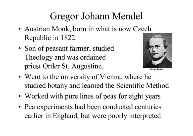
Gregor Johann Mendel
• Austrian Monk, born in what is now Czech
Republic in 1822
• Son of peasant farmer, studied
Theology and was ordained
priest Order St. Augustine.
• Went to the university of Vienna, where he
studied botany and learned the Scientific Method
• Worked with pure lines of peas for eight years
• Pea experiments had been conducted centuries
earlier in England, but were poorly interpreted
• Conducted pea research between 1856 and
1863
• In 1866 he published Experiments in Plant
Hybridization, (Versuche über PflanzenHybriden) in which he
established his three
Principles of Inheritance
• Work was largely ignored for
34 years, until 1900, when
3 independent botanists
rediscovered Mendel’s work.
(De Vries, von Tschermak & Correns)
• Mendel was the first biologist to use
mathematics to explain his results
quantitatively.
• Mendel predicted
– The concept of genes
– That genes occur in pairs
– That one gene of each pair is present in the
gametes
Genetics terms you need to
know:
• Gene – a unit of heredity;
a section of DNA sequence
encoding a single protein
• Genome – the entire set
of genes in an organism
• Alleles – two genes that occupy the same
position on homologous chromosomes and
that cover the same trait (like ‘flavors’ of a
trait).
• Locus – a fixed location on a strand of DNA
where a gene or one of its alleles is located.
• Homozygous – having identical genes (one
from each parent) for a particular
characteristic.
• Heterozygous – having two different genes
for a particular characteristic.
• Dominant – the allele of a gene that masks
or suppresses the expression of an alternate
allele; the trait appears in the heterozygous
condition.
• Recessive – an allele that is masked by a
dominant allele; does not appear in the
heterozygous condition, only in
homozygous.
• Genotype – the genetic makeup of an
organisms
• Phenotype – the physical appearance
of an organism (Genotype + environment)
• Monohybrid cross: a genetic cross
involving a single pair of genes (one trait);
parents differ by a single trait.
• P = Parental generation
• F1 = First filial generation; offspring from a
genetic cross.
• F2 = Second filial generation of a genetic
cross
Mendel’s Principles
• 1. Principle of Dominance:
One allele masked another, one allele
was dominant over the other in the F1
generation.
• 2. Principle of Segregation:
When gametes are formed, the pairs
of hereditary factors (genes) become
separated, so that each sex cell
(egg/sperm) receives only one kind of
gene.
Principle of Independent
Assortment
Based on the pea results, Mendel postulated
the
3. Principle of Independent Assortment:
“Members of one gene pair segregate
independently from other gene pairs during
gamete formation”
Genes get shuffled – these many combinations
are one of the advantages of sexual
reproduction
A Warning on Assortment
• Today we know independent
assortment works only if the genes lie
on different chromosomes
• If two genes lie on the same
chromosome, they will be transmitted
together
• Mendel looked at seven traits he
reported as independently assorted.
His peas had seven pairs of
chromosomes. Historians say he likely
threw away data that did not fit his
hypotheses!
Coming Next:
•
•
•
•
Incomplete dominance
Human blood types
Complex traits
Sex-linked traits
Dominanza incompleta o codominanza
Quando nell’eterozigote i due alleli si esprimono entrambi in egual
misura e l’espressione di ciascun allele è riconoscibile a livello
fenotipico.
Esempio: gruppi sanguigni sistema ABO
Genotipo
IA – IA
IA – i
IB – IB
IB – i
IA – IB
i-i
Fenotipo
Gruppo A
Gruppo B
Gruppo AB
Gruppo 0
Penetranza: la probabilità che un allele si esprima negli individui che lo
possiedono;
Penetranza completa: se il 100% degli individui portanti un determinato allele
manifestano il fenotipo corrispondente;
Penetranza ridotta o incompleta: se la frequenza di espressione è inferiore al
100%.
Espressività: grado di manifestazione del carattere;
Espressività uniforme: il carattere fenotipico è uguale in tutti gli individui
Espressività variabile: manifestazione fenotipica differente in individui con lo
stesso genotipo;
:
Geni localizzati sul cromosoma X
La trasmissione ereditaria dei geni X-linked è diversa da quella dei geni
autosomici in quanto si osserva differenza tra gli incroci reciproci, cioè
la F1 è diversa a seconda che un carattere sia trasmesso dal padre o
dalla madre.
Punnett Square for Sex Determination
Reginald Punnett
(1875-1967) developed
this device to explain
sex determination. He
explored sex-linked
coloration in chickens.
Female gametes across top
Male gametes along side
Inattivazione del cromosoma X nelle cellule di mammifero
Modelli di Ereditarietà
Autosomica
Il gene la cui mutazione è responsabile dell’insorgenza del fenotipo è
localizzato sugli autosomi
X-linked
Il gene è localizzato sul cromosoma X (differente l’espressione nei due
sessi)
Y-linked
Il gene è localizzato sul cromosoma Y (Eredità paterna)
Mitocodriale
Il fenotipo è determinato da geni localizzati nel genoma mitocondriale
(Eredità materna)
Modelli di Ereditarietà
Dominante
L’eterozigote manifesta il fenotipo (guadagno di funzione)
Recessivo
Soltanto l’omozigote manifesta il fenotipo (perdita di funzione)
Autosomica Dominante
Neurofibromatosi
Corea di Huntington
Autosomica Recessiva
Talassemie
Falcemia
Fibrosi cistica
Fenilchetonuria
X-linked
Distrofia muscolare
Cecità ai colori
Favismo
X-fragile
Year
1986
Disease
Duchenne muscular
dystrophy
MIM n
310200
Location Gene
Xp21.3 DMD
Chromosome abnormality
(a) del(X)(p21.3)
1989
1990
Retinoblastoma
Cystic fibrosis
Neurofibromatosis 1
180200
219700
162200
13q14
7q31
17q11.2
RB
CFTR
NF1
1991
Wilms' tumor
Aniridia
194070
106210
11p13
11p13
WT1
PAX6
Familial polyposis coli
Fragile-X syndrome
Myotonic dystrophy
Huntington's disease
Tuberous sclerosis 2
175100
309550
160900
143100
191092
5q21
Xq27.3
19q13.3
4p16
16p13
APC
FMR1
DMPK
HD
TSC2
von Hippel-Lindau disease 193300
3p25
VHL
Achondroplasia
100800
Early-onset breast/ovarian 113705
cancer
Polycystic kidney disease 173900
601313
Spinal muscular atrophy 253300
600354
4p16
17q21
FGFR3
BRCA1
(b) t(X;21)(p21.3:p13)
del(13)(q13.1q14.5)
None
Balanced translocations
t(1;17)(p34.3:q11.2)
t(17;22)(q11.2:q11.2)
del(11)(p14p13)
t(4;11)(q22;p13)
del(11)(p13)
del(5)(q15q22)
FRAXA fragile site
None
None
Microdeletions in candidate
region
Microdeletions in candidate
region
None
None
16p13.3
PKD1
t(16;22) (p13.3;q11.21)
5q13
SMN1
None
1993
1994
1995
OMIM Statistics for May 26, 2008
Autosomal
X-Linked
Y-Linked
Mitochondrial
Total
Gene with known
sequence
11686
538
48
37
12309
Gene with known
sequence
and phenotype
358
30
0
0
388
Phenotype
description,
molecular basis
known
2101
196
2
26
2325
Mendelian phenoty
pe or locus,
molecular basis
unknown
1477
136
5
0
1618
Other, mainly
phenotypes with
suspected mendelian
basis
1944
140
2
0
2086
17566
1040
57
63
18726
*
+
#
%
Total
•(A) Autosomal dominant;
•(B) autosomal recessive;
•(C) X-linked recessive;
•(D) X-linked dominant;
•(E) Y-linked.
04_02.jpg
Genoma Mitocondriale
16.600 bp
37 geni
Eredità mitondriale
o
Eredità materna
LEBER HEREDITARY OPTIC NEUROPATHY; LHON
Human case: CF
• Mendel’s Principles of Heredity apply
universally to all organisms.
• Cystic Fibrosis: a lethal genetic disease
affecting Caucasians.
• Caused by mutant recessive gene carried by
1 in 20 people of European descent (12M)
• One in 400 Caucasian couples will be both
carriers of CF – 1 in 4 children will have it.
• CF disease affects transport
in tissues – mucus is accumulated
in lungs, causing infections.
Inheritance pattern of CF
IF two parents carry the recessive gene of
Cystic Fibrosis (c), that is, they are
heterozygous (C c), one in four of their
children is expected to be homozygous
for cf and have the disease:
C
C C = normal
C c = carrier, no symptoms
c c = has cystic fibrosis
c
C
CC
Cc
c
Cc
cc
Neuropsychiatric diseases caused by expansion
of trinucleotide repeats
• Myotonic dystrophy
• Fragile X syndrome
• Spinal and bulbar muscular atrophy (Kennedy’s)
• Huntington’s disease
Microsatellites
• short regions of repeating DNA sequence in the genome
(because their G+C content is usually higher or lower
than the average for the genome they frequently appear
to band at a different buoyant density in CsCl gradients
and hence are called “satellites”)
• microsatellites are often comprised of “trinucleotide repeats”
X fragile
(309550)
Frequenza: 1/4000 maschi.
Ereditarietà: Legata al cromosoma X. Malattia causata da mutazione
dinamica.
Genetica: Nel 1991 è stato identificato il gene responsabile. La
mutazione è caratterizzata dall’amplificazione di un tratto di DNA
costituito da una specifica sequenza ripetuta (CGG). Nei soggetti
normali è presente un numero di ripetizioni variabili da 6 a 55.
Esistono due differenti tipi di mutazione: la premutazione (56-200) e
la mutazione completa (>200). La probabilità di espansione aumenta
con le dimensioni della premutazione e quindi con il passare delle
generazioni (Paradosso di Sherman).
Diagnosi: La diagnosi molecolare (Southern blot) permette di
individuare anche gli individui con la premutazione.
Malattia di Huntington
(143100)
Frequenza: 5-10/100.000 nati vivi
Ereditarietà: autosomica dominante. Malattia causata da mutazione
dinamica
Genetica: Il gene responsabile della malattia ed il suo prodotto proteico
sono stati identificati. Il gene definito Intersting Transcript (IT-15), è
localizzato sul braccio corto del cromosoma 4 (4p16.3). La malattia è
associata all’amplificazione patologica di una specifica sequenza ripetuta
(CAG) nell’allele mutato. Nella popolazione normale la tripletta è ripetuta
10-30 volte. Nei pazienti affetti il numero di ripetizioni varia da 36 a più di
100. Un numero intermedio di espansioni 30-35 volte, è considerato una
premutazione.
Diagnosi: Il test genetico si basa sulla determinazione del numero di
espansione della tripletta.
Human Genome Project
1990 – 2001 – ………….
15 February 2001
Studio delle malattie genetiche
Classificazione
????????????
Numero di geni
Malattie monogeniche e Fenotipi complessi
Tipo cellulare
Zigote o mutazioni somatiche
Funzione del gene interessato
La mutazione è presente
Malattie congenite nello zigote
Malattie ad
esordio tardivo
La mutazione (o le
mutazioni) insorge durante
la vita dell’individuo in
cellule somatiche
Mutazioni somatiche e germinali
Malattie congenite
•Mutazioni geniche
•Alterazioni complesse del genoma
(mutazioni cromosomiche)
Malattie ad esordio tardivo
•Cancro
•Malattie degenerative
•Malattie infettive
Presenti nello zigote
Alterazioni complesse del genoma
Mutazioni cromosomiche
This database is a catalog of human genes and genetic disorders authored and
edited by Dr. Victor A. McKusick and his colleagues at Johns Hopkins and
elsewhere,
and developed for the World Wide Web by NCBI, the National Center for
Biotechnology Information.
The database contains textual information and references.
It also contains copious links to MEDLINE and sequence records in the Entrez
system, and links to additional related resources at NCBI and elsewhere.
http://www.ncbi.nlm.nih.gov/entrez/query.fcgi?db=OMIM
Studio delle malattie genetiche
Dal Fenotipo al Genotipo
Dal Genotipo al Fenotipo
Studio delle malattie genetiche
Dal Fenotipo al Genotipo
Dal Genotipo al Fenotipo
Commonly used methods for identifying genes in cloned DNA
Method
Zoo blotting
Comments
A DNA clone is hybridized at reduced hybridization stringency against a
Southern blot of genomic DNA samples from a variety of animal species, a
zoo blot.
Depends on coding DNA being more strongly conserved in evolution than
non-coding DNA (Figure 10.21).
Many vertebrate genes have associated CpG islands, hypomethylated GCrich sequences usually having multiple rare-cutter restriction sites ( Cross
and Bird, 1995).
CpG island identification
Identification by restriction mapping. DNA clones are usually hybridized
against Southern blots of genomic DNA cut with SacII, EagI or BssHII to
identify clustering of rare-cutter sites (Figure 10.22).
Island-rescue PCR. This is a way of isolating CpG island sequences from
YACs by amplifying sequences between islands and neighbouring Alu
repeats.
Hybridization
A genomic DNA clone can be hybridized against a Northern to mRNA/cDNA
blot of mRNA from a panel of culture cell lines, or against appropriate cDNA
libraries.
Exon trapping
This is essentially an artificial RNA splicing assay (see Figure 10.23). It relies
on the observation that the vast majority of mammalian genes contain
multiple exons which need to be spliced together at the RNA level.
cDNA selection or capture
These techniques involve repeated purification of a subset of genomic DNA
clones which hybridize to a given cDNA population (see Figure 10.24).
Computer analysis of DNA
sequence
Homology searches. Any DNA sequence obtained from a genomic clone can
be compared against all other sequences in sequence data-bases. Significant
homology to known coding DNA or gene-associated sequences may indicate
a gene (see Section 20.1.4)
Gene searching algorithms. A variety of computer programs have been
developed to search sequences for exons and other gene-associated motifs
(see Figure 10.25 and Section 20.1.4).
Year
1986
Disease
Duchenne muscular
dystrophy
MIM n
310200
Location Gene
Xp21.3 DMD
Chromosome abnormality
(a) del(X)(p21.3)
1989
1990
Retinoblastoma
Cystic fibrosis
Neurofibromatosis 1
180200
219700
162200
13q14
7q31
17q11.2
RB
CFTR
NF1
1991
Wilms' tumor
Aniridia
194070
106210
11p13
11p13
WT1
PAX6
Familial polyposis coli
Fragile-X syndrome
Myotonic dystrophy
Huntington's disease
Tuberous sclerosis 2
175100
309550
160900
143100
191092
5q21
Xq27.3
19q13.3
4p16
16p13
APC
FMR1
DMPK
HD
TSC2
von Hippel-Lindau disease 193300
3p25
VHL
Achondroplasia
100800
Early-onset breast/ovarian 113705
cancer
Polycystic kidney disease 173900
601313
Spinal muscular atrophy 253300
600354
4p16
17q21
FGFR3
BRCA1
(b) t(X;21)(p21.3:p13)
del(13)(q13.1q14.5)
None
Balanced translocations
t(1;17)(p34.3:q11.2)
t(17;22)(q11.2:q11.2)
del(11)(p14p13)
t(4;11)(q22;p13)
del(11)(p13)
del(5)(q15q22)
FRAXA fragile site
None
None
Microdeletions in candidate
region
Microdeletions in candidate
region
None
None
16p13.3
PKD1
t(16;22) (p13.3;q11.21)
5q13
SMN1
None
1993
1994
1995
Human Genome Project
1990 – 2001 – ………..
Studio delle malattie genetiche
Diagnosi
Cura
Prevenzione
Multiplex ARMS test per individuare 4
specifiche mutazioni che causano la fibrosi
cistica
Diagnosi di malattie da
espansione di triplette ripetute
MEDICINA MOLECOLARE

